Project background
In the EU, two forms of echinococcosis are clinically recognized in humans: Cystic Echinococcosis (CE), caused by Echinococcus granulosus complex and Alveolar Echinococcosis caused by Echinococcus multilocularis. However, human CE is the most prevalent and probably accounts for more than 95% of the estimated 2-3 million global cases with 200,000 new cases diagnosed annually. CE is one of the most widespread zoonotic diseases, although not evenly distributed geographically.
In the North and Western Europe few cases of CE are detected in livestock and most human cases are imported. On the contrary, in many parts of the world, CE causes severe healthcare and economic losses due to treatment costs, lost wages, and livestock-associated production losses.
In the European Union and adjacent countries, some southern countries (such as Spain and Italy) and eastern countries (such as Bulgaria, Romania and Turkey) are those where human CE is highly endemic and it is still a major health and economic problem. In Eastern Europe, the number of CE human cases has dramatically increased in recent years, with high endemicity in pastoral communities. For instance, an ultrasound screening survey conducted by members of WHO-IWGE (WHO Informal Working Group on Echinococcosis) on 4,146 subjects living in rural areas of Ialomita, Buzau and Teleorman counties in Romania during the period 2005-2009, detected 173 people with CE with an impressive prevalence between 3.7 and 4.9%.
The results from HERACLES will support governments, the European Commission and related European agencies such as ECDC to harmonize data collection, monitoring and reporting of CE, according to the EU legislation.
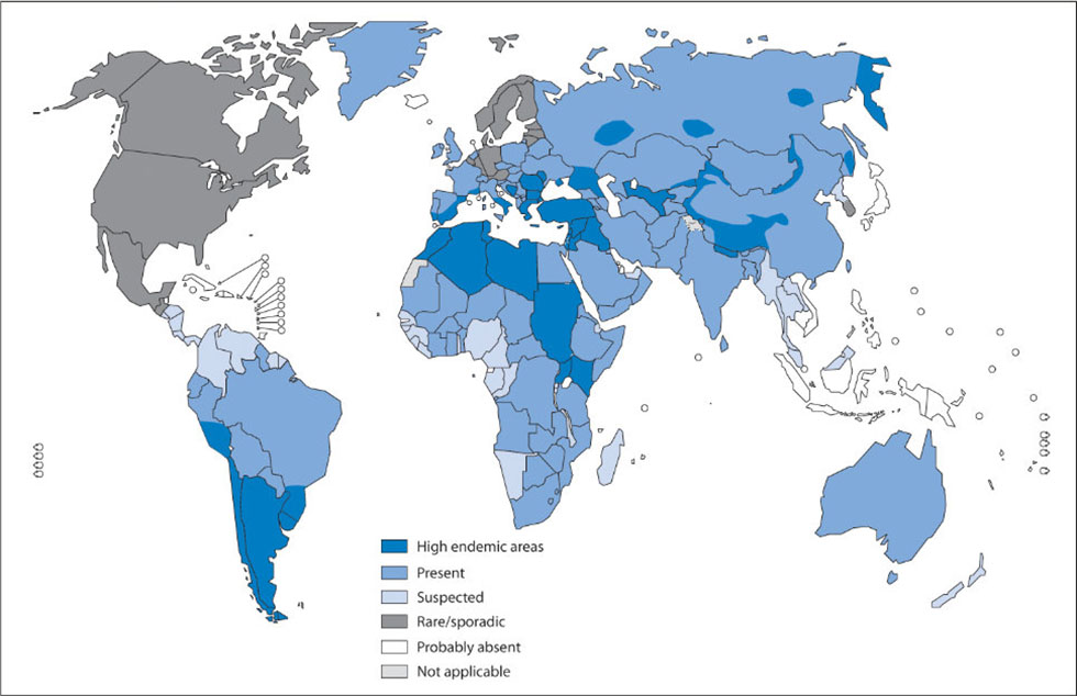
Global distribution of cystic echinococcosis. Italy, Spain and Eastern European countries (such as Bulgaria, Romania and Turkey) are recognized as highly endemic (from WHO web-page, Global Health Observatory Map Gallery).
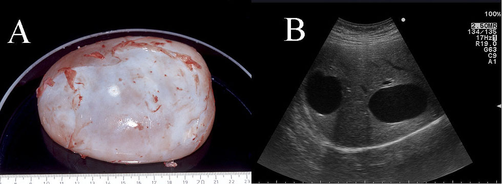
A. Intact larval part of CE1 echinococcal parasitic cyst in a Petri dish after surgery (courtesy Prof. Hans Seitz, University of Bonn, Bonn, Germany); B. Two CE1 cysts stages seen on ultrasound in the right hepatic lobe. Fluid is anechogenic (black) on ultrasound.